

What is Megaesophagus?
Megaesophagus is a disease that affects Friesian horses and is characterized by chronic dilation of the esophagus. The prevalence of the disease in Friesian horses and the very young age at which some Friesians are affected suggests a genetic inheritance.
Megaesophagus has varying degrees of severity. Symptoms may initially be very mild and challenging for owners to identify. As the disease progresses, dilation of the esophagus occurs, most frequently in the thoracic region of the esophagus, often near the thoracic inlet. However, in some cases, the entire esophagus may be affected, or the cervical portion may be affected.
If the disease goes unnoticed, severe complications can occur, including esophageal ulcers, perforations, and strictures. Eventually, if no intervention is made, the esophagus can rupture.
Unfortunately, there is no treatment or medication for megaesophagus. Diet management is the most useful approach.
Numerous research studies and the data collected by the Fenway Foundation support the theory that the most likely cause of megaesophagus in Friesians is a connective tissue disorder. However, research is ongoing.
If your Friesian horse has been diagnosed with megaesophagus or you suspect they have megaesophagus, please consider supporting our research by submitting a hair sample.
Connective Tissue

Structure of the esophagus

Most Friesians are affected in the thoracic region of the esophagus

Flehmen-like response

Latherin (white foam) while eating

Choke with reflux

Grazing reflux
Symptoms of Megaesophagus
-
Repeated episodes of choke
-
Hypersalivation (drool)
-
Aspiration pneumonia
-
Difficulty swallowing
-
Weight loss
-
Ulcers
-
Rolling
-
Pawing
-
Bad breath
-
Head shaking
-
Teeth grinding
-
Excess mucous
-
Nasal discharge
-
Teeth chomping
-
Excessive licking
-
Refusing feed or hay
-
Sticking tongue out
-
Colic-like symptoms
-
Moving tongue around
-
Standing with head down
-
Heavy/labored breathing
-
Lowering or extending the neck
-
Appearing anxious or nervous
-
Standing alone, unusually quiet
-
Repeated coughing or snorting
-
Milk coming out the nose in foals
-
Dramatic coughing during exercise
-
White foam around or in the mouth
-
Gurgling or gagging noises while eating
-
Repeated lowering and raising of the head
-
Loss of pigmentation on the edge of the mouth
-
Standing stretched out with head high in the air
-
Throat clearing or pop-like noise when swallowing
-
Standing with front legs spread wider than normal
-
Flehmen-like response (lifting/curling upper lip)
-
Excessive coughing, sneezing, or saliva while exercising
-
Wave-like motion of the esophagus while swallowing
-
Visible firm bulge of the esophagus near the base of the chest
-
Profuse reflux of feed material through the mouth and/or nose
Diagnosing Megaesophagus
Megaesophagus is typically diagnosed by performing an esophagoscopy, commonly referred to as a "scope", or a barium swallow study.
Esophagoscopy
An esophagoscopy is an examination of the esophagus using a thin, tube-like scope with a light and a camera. An esophagoscopy allows the veterinarian to visualize the esophagus and look for dilated areas, poor motility, and complications such as strictures, ulcers, or diverticula. The downside of an esophagoscopy is that it is subjective and relies on the observations and experience of the veterinarian performing it for an accurate diagnosis. In some rural areas, a veterinarian might have never seen megaesophagus, and in some cases, the esophagus is only mildly affected. The Fenway Foundation has seen dozens of cases where a Friesian was initially cleared of megaesophagus after an esophagoscopy, only to be diagnosed with megaesophagus later following a barium swallow study.
Barium Swallow Study
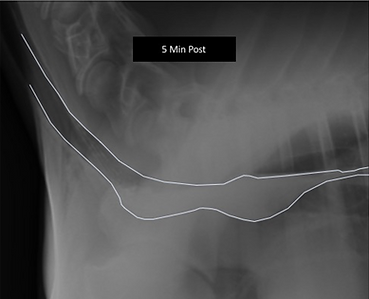
A barium swallow study is a key diagnostic tool for evaluating megaesophagus, and it involves administering a chalky, white substance orally called "barium". The barium coats the surface of the esophagus and appears white on radiographs. The procedure produces detailed images of the linings of the esophagus. It can provide insight into the motility of the horse's swallowing and the location of any dilated portions of the esophagus.
Recommendations
The Fenway Foundation recommends both an esophagoscopy and a barium swallow study to definitively diagnose megaesophagus. Despite some of its drawbacks, an esophagoscopy is a critical diagnostic for megaesophagus, as a barium swallow study alone cannot visualize the health of the esophageal tissue and identify all complications. By combining both diagnostic tools, owners can ensure their Friesian receives a thorough and accurate diagnosis
Images from a esophagoscopy

Images from a barium swallow study


Managing
Megaesophagus
Megaesophagus is a disease that affects Friesian horses and is characterized by chronic dilation of the esophagus. The prevalence of the disease in Friesian horses and the very young age at which some Friesians are affected suggests a genetic inheritance.
Megaesophagus has varying degrees of severity. Symptoms may initially be very mild and challenging for owners to identify. As the disease progresses, dilation of the esophagus occurs, most frequently in the thoracic region of the esophagus, often near the thoracic inlet. However, in some cases, the entire esophagus may be affected, or rarely, only the cervical portion may be affected.
Horses with megaesophagus can be managed with a strict diet and management protocol, but the prognosis is guarded to poor. We assist owners all over the world with diet and management plans for horses with megaesophagus. Please reach out to us if you would like a consult.
Below are basic diet recommendations for horses with megaesophagus. Depending on the severity of a horse's disease and complications (esophageal diverticula, esophageal ulcers, esophageal strictures, esophagitis, etc.), they may require a more restrictive diet.


Community Support

For more information or community support, please join our Facebook support group, Equine Megaesophagus.